


Varicose veins during pregnancyare ectases of venous blood vessels that occur during pregnancy and are pathogenetically associated with it. Its severity, paraesthesia, pain in the lower extremities and external genitalia, swelling, muscle twitching, trophic skin lesions are manifested. It is diagnosed by examination with ultrasound angioscanning methods. During pregnancy, treatment is usually limited to compression therapy with correction of sleep and rest, physical activity, and nutrition. Perhaps the appointment of phlebotonics, phleboprotectors, anticoagulants, platelet inhibitors. Surgical treatments are usually used after childbirth.
General Information
Varicose veins (varicose veins) are one of the most common vascular diseases associated with pregnancy. Studies show that up to 15-20% of people suffer from venous pathology, while 2/3 are female, and 60-80% of venous ectasia cases are due to pregnancy. The disease is usually diagnosed first in young patients, 75% of whom are under 30 years of age. In more than two-thirds of cases, varicose veins debut after the 20th week of their first pregnancy. The relevance of early diagnosis of varicose veins is associated with a high probability of fetoplacental insufficiency and a risk of fatal thromboembolic complications in the absence of appropriate therapy.
Causes
Based on statistics on the incidence of varicose veins during pregnancy, most obstetricians and gynecologists consider the disease to be a complication of pregnancy. The predisposing factor for vascular ectasis in 91% of patients is genetically determined failure of the middle venous sheath, in which the amount of collagen material decreases and the content of polysaccharides increases. The development of varicose veins in constitutionally predisposed women during pregnancy is aided by:
- Increased circulating blood volume. The increase in BCC in pregnant women ranges from 30-50% (when carrying 1 child) to 45-70% (when there are 2 or more fetuses in the womb). This compensation mechanism allows the child to have an adequate blood supply, the woman’s vital organs, and the fetoplacental system.
- Hormonal adjustment during pregnancy. During pregnancy, the ovaries and placenta excrete progesterone and relaxin intensively. As a result of these hormones, the smooth muscle fibers of the veins relax and structural reconstruction of the connective tissue occurs. As a result, the vessel wall copes worse with increased intravenous pressure.
- Compression of blood vessels by the pregnant uterus. The growing uterus compresses the inferior vena cava and iliac veins. Blood flows out of the pelvis and lower extremities, increasing intravascular pressure, which provokes stretching of the venous walls. The effect of this factor plays a key role in the development of varicose veins after the 25th week of pregnancy.
- Changes in the hemostatic system. As labor approaches, blood fibrinolytic activity decreases and the number of coagulation factors increases. The purpose of this adaptation mechanism is to reduce the amount of physiological blood loss during labor. This increases the likelihood of thrombosis of abnormally altered veins.
Another etiofactor that contributes to the development of varicose veins in pregnant women is a decrease in physical activity. Insufficient work of skeletal muscles increases blood stagnation in the legs and pelvis. The situation is exacerbated in the presence of obesity, when the amount of blood circulating in the patient's vascular bed increases even more.
Pathogenesis
The starting point for the development of varicose veins during pregnancy is disruption of the compensatory capabilities of the venous valve device. Due to the increase in BCC and the mechanical blockage of the outflow from the lower extremities, the blood exerts increased pressure on the vessel wall when the main veins are constricted. Genetically inherited connective tissue insufficiency is enhanced by relaxation of vascular smooth muscle under the influence of progesterone. As a result, the lumen of the vein dilates, the valves stop, and blood is deposited in the vascular system of the lower limbs. As the disease progresses, the pathological process can spread to the vessels of the vulvar ring, vagina, and small pelvis.
Classification
The main criteria for systematizing varicose veins are the anatomical prevalence of venous congestion and the severity of the disease. This approach allows for the differentiated selection of disorders for different variants of the disorder. Considering the involvement of different organs in the process, we distinguish between varicose veins of the lower limbs, varicose veins, and varicose veins of the pelvic organs. According to the severity of the clinical symptoms, the following stages of dilation of the venous vessel in the lower extremities are distinguished:
- Compensated varicose veins. There are no external signs of vascular ectasia, the pregnant woman notes by the end of the day the fatigue of the legs, the discomfort of the calf muscles during exercise and rapid walking.
- Subcompensated varicose veins. Vascular bowel ("stars") appears on the skin. In the evening the legs swell, at night cramps, numbness, pain occur. Bruises and scratches heal further than usual.
- Decompensated varicose veins. The patient is constantly worried about the pain in the legs, the swelling is increasing. The blood vessels are significantly enlarged, lumpy. The skin is hyperpigmented. There are signs of eczema and trophic disorders.
In the case of pelvic varicose veins, the disease also develops intermittently. In the first stage, the diameter of the affected vessels in any venous plexus of the pelvis does not exceed 5. 0 mm. With the second, the uterus or ovary is involved in the process, the lumen of the blood vessels is 6, 0-10, 0 mm. The third is characterized by ectasia of veins larger than 10 mm, causing complete damage to all venous plexuses in the pelvis.
Symptoms of varicose veins
In 80-82% of patients, the disease debuts with a feeling of heaviness and tension, the legs "buzz", intensifying in the evenings and during physical exertion. The symptoms of varicose veins gradually increase. As the disease progresses, there is pain in some areas of the muscles, which first ends with prolonged work and physical work. In the most severe cases, the pain becomes constant, its intensity can be so pronounced that the pregnant woman has difficulty moving independently. 60% of patients notice cramps in the calf muscles, up to 40-50% - loss of sensitivity, numbness of the legs, up to 30% - itching.
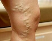
In the subcompensated stage of the varicose veins, external signs of dilation of the superficial veins appear. First, areas of reticular vessels and telangiectasias ("reticules" and "stars") form on the skin. Thereafter, the venous pattern becomes clear. The veins appear dilated, twisted, and eventually nodular. The spread of the process of ectasia to the deep blood vessels is evidenced by the occurrence of edema in the ankle joints and lower legs. With the decompensation of the varicose veins, the skin of the legs appears hyperpigmented, eczema develops. If the pathology occurred long before pregnancy, dystrophy of the subcutaneous adipose tissue, trophic ulcers, is possible.
In 4% of patients, the disease affects the veins of the vulva, vagina and pelvis. In the case of vulvar and vaginal varicose veins, discomfort, swelling, heaviness, itching can be observed in the external genital area. Swelling of the perineum and labia, contact bleeding from the vagina after sex may occur. Pelvic congestion syndrome is manifested by pain in the lower abdomen or painful pain that radiates to the lower back, sacrum, groin, and external genitals. Dyspareunia (pain during intercourse) is typical. In severe cases, dysuric disorders are observed.
Complications
In the absence of appropriate treatment, varicose veins in pregnant women can be complicated during trophic ulcers, erysipelas, thrombophlebitis, superficial and deep vein thrombosis, pulmonary artery, and other large vessel thromboembolism during labor. In 40-45% of cases, placental insufficiency occurs with acute and chronic fetal hypoxia. Workforce disorders (weakness of the workforce, discoordination of myometrial contractile activity) are observed in 25% of patients. In the case of vaginal varicose veins, massive traumatic course of the postpartum period is possible. Nearly one-third of women in labor have defects in placental abruption and placental emptying. The long-term consequences of varicose veins during pregnancy are hemorrhoids, chronic venous insufficiency, and pelvic pain.
Diagnostics
With the appearance of characteristic skin marks, the diagnosis of varicose veins during pregnancy usually does not cause difficulties. The task of the diagnostic phase is to determine the stage and localization of venous ectasia, to rule out other causes that may cause stagnation in the vascular system of the lower extremities. The most informative survey methods are:
- Presidential Review. The study reveals the characteristic changes of the venous vessels in the vulvar region and the inner thighs - ectasia, tortuity, nodosity. Swelling of the labia and perineum is possible. Seen from the mirrors, the vaginal mucosa appears hypertrophied, cyanotic. Bimanual tactile vaginal vaults are smooth, often painful.
- the venous system is USDG. Ultrasound examines the shape and diameter, length, anatomical position, and wall condition of the blood vessels. The method allows the determination of branching zones, consistency of valve equipment, permeability of veins, presence and direction of reflux. You can scan the veins in your lower limbs and lower vena cava (IVC ultrasound).
- Duplex scanning of footers. The advantage of the non-invasive method, which combines traditional ultrasound and Doppler examinations, is not only to obtain detailed information on blood flow parameters, but also to visualize the venous network. Duplex angioscanning is used to comprehensively assess the condition of surface, perforating, and deep vessels.
Radiodiagnostic methods during pregnancy (varicography, selective ovarian uptake, ascending phlebography of the extremities, pelvic phlebography, CT venography, phleboscintigraphy, etc. ) are used to a limited extent due to possible adverse effects on the fetus. In severe cases, with suspected pelvic varicose veins, diagnostic laparoscopy should be performed with caution. Differential diagnosis of leg varicose veins is performed with drops in pregnant women, heart failure, lymphedema, and acute thrombosis of the venous system. Varicose veins in the pelvis should be distinguished from genital endometriosis, chronic inflammatory pathology of the pelvic organs, submucosal and suberous uterine fibroids, cysts, and other ovarian tumors. In addition to being monitored by an obstetrician-gynecologist, the patient is advised to consult a phlebologist, cardiologist, and oncologist.
Treating varicose veins during pregnancy
The main goal of varicose vein therapy in pregnant women is to arrest the progression of the disorder, alleviate the severity of the clinical picture, and prevent possible thromboembolic complications. Non-pharmacological methods are preferred, supplemented, if necessary, by drug therapy during the safe period of pregnancy:
- Compression therapy. A woman with a confirmed diagnosis of varicose veins is recommended to wear daily during pregnancy, elastic bandage, special compression stockings or stockings of compression class 1-2 in the postpartum and postpartum period. Compression treatment accelerates blood flow, reduces swelling and congestion by mechanically reducing the diameter of the superficial veins.
- Herbal phlebotonics and phleboprotectors. The effect of the use of drugs belonging to this group is associated with an increase in the tone of the venous wall, a decrease in its permeability, an improvement in microcirculation, and an improvement in the rheological properties of blood and lymph outflow. The advantage of most bioflavonoids is that they can be used during pregnancy and lactation. Phlebotonic drugs are prescribed both in tablet form and externally.
- Anticoagulants and platelet inhibitors. Drugs with antithrombotic activity should be used with caution in the presence of signs of an increased tendency to coagulation and the risk of developing DIC. In order to improve the rheology of blood and vascular microcirculation, pharmaceutical compositions that prevent platelet aggregation and exert an angioprotective effect are presented.
Special physiotherapy exercises, lymphatic drainage massage, dosing walk, daily rising contrast shower are recommended for varicose pregnant women. Adjusting your diet means eating foods rich in fiber and vegetable fats. Injectable sclerotherapy, miniflebectomy, crossectomy, endovascular laser coagulation, and other surgical methods are used in exceptional cases with severe forms of the disease, severe pain syndrome, and the presence of complications. Most often, surgical correction is performed at the end of the lactation period.
Shipping Tactics
The preferred mode of transport for varicose veins is natural childbirth, which begins with the use of elastic bandages or compression garments on the lower extremities of the woman in labor. In patients with vulvar-vaginal varicose veins, the long-term period requires particularly careful support, with protective perineotomy, as indicated. When ectasized veins rupture, the damaged vessels are carefully connected by re-suturing the conglomerate of nodes. Cesarean section is recommended for patients at high risk of thromboembolic complications and severe varicose veins.
Forecasting and prevention
With timely detection and appropriate therapy, the prognosis is favorable. As a preventative measure, adequate nighttime sleep and periodic rest are recommended in a supine position during the day with the legs lying on a solid surface at an angle of 30 °. Pregnant women with an inherited inheritance should not refuse to wear shoes with heels larger than 5 cm, limit the duration of sitting or standing, and monitor weight gain.
Daily walks, reduced salt intake and vitamin wall-strengthening vitamin preparations are effective in preventing varicose veins. Varicose vein patients planning a pregnancy are indicated to undergo surgical interventions to correct the disease.